An AI system can diagnose brain tumor in 150 seconds
7. Januar 2020An AI system can diagnose brain tumor in 150 seconds
New York, January 7, 2020
Human pathologists need about 30 minutes to diagnose brain tumors from tissue samples taken during surgery. A new AI system can do this in less than 150 seconds – and with greater accuracy than its human counterparts, according to research recently published in Nature Medicine.
It describes a novel diagnostic procedure that combines the power of artificial intelligence with an advanced optical imaging method. The system is able to make fast and accurate diagnoses of brain tumours in virtually real time while the patient is still lying on the operating table. In tests, the AI has made diagnoses in a fraction of the time that were slightly more accurate than those made by human pathologists. It is exciting that the new system could be used in areas where neurological experts are not available, and it is a promising technique that could also diagnose other types of cancer, including dermatology, gynecology, breast surgery and head and neck surgery.
In cancer surgery, it is not uncommon for surgeons to remove potentially problematic tissue for laboratory analysis. These intraoperative biopsies allow more accurate diagnoses and help the medical team plan the next steps, such as planning a subsequent operation to remove the tumor.
According to the new study, around 1.1 million brain samples are biopsied in the USA every year – all of them are meticulously examined by a trained neuropathologist. This process is, as the authors write in the paper, „time, resource and labour intensive“.
In fact, these diagnoses involve more than a dozen steps, including transporting the tissue from the operating theatre to the laboratory and temporarily freezing it, thawing and dehydrating the sample, cleaning it with xylene and placing it under a microscope for analysis – not to mention all the steps the pathologist has to take to assess the tissue. The situation is further complicated by the fact that there is currently a shortage of neuropathologists in the USA.
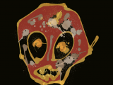
To streamline this process, neuroscientist Daniel Orringer and his colleagues at New York University developed a diagnostic method that combines a powerful new optical imaging technique known as stimulated Raman histology (SRH) with an artificially intelligent deep neural network. SRH uses scattered laser light to illuminate features that are normally not visible in standard imaging techniques. During surgery, the images acquired by SRH are evaluated by the AI algorithm, which takes less than 150 seconds to evaluate, compared to the 20 to 30 minutes required by human neuropathologists. The authors „demonstrated how the combination of SRH with deep learning can be used to rapidly predict intraoperative brain tumor diagnosis. It is fascinating that AI is also able to detect features in biopsies that are not visible to the human eye.
„As surgeons, we are limited to responding to what we can see; this technology allows us to see what would otherwise be invisible to improve speed and accuracy in [the operating room] and reduce the risk of misdiagnosis,“ Orringer, the senior author of the paper, said in a press release. „With this imaging technology, cancer surgery is safer and more effective than ever before.“
To create the deep neural network, the scientists trained the system on 2.5 million images from 415 patients. At the end of the training, the AI was able to classify the tissue into 13 common types of brain tumors, including malignant glioma, lymphoma, metastatic tumors, diffuse astrocytoma and meningioma.
A clinical trial involving 278 brain tumour and epilepsy patients and three different medical institutions was then set up to test the effectiveness of the system. The SRH images were evaluated either by human experts or the AI. The results showed that the AI correctly identified the tumor in 94.6 percent of the cases, while the neuropathologists were correct in 93.9 percent of the cases. Interestingly, the human errors were different from the errors of the AI. This is actually good news because it suggests that the nature of the AI’s mistakes can be taken into account and corrected in the future, which will lead to an even more accurate system, the authors say.
„SRH will revolutionize the field of neuropathology by improving decision making during surgery and enabling expert level assessment in hospitals where trained neuropathologists are not available,“ said Matija Snuderl, a co-author of the study and associate professor at the NYU Grossman School of Medicine.